


")


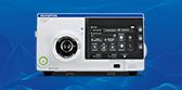
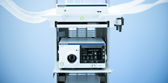
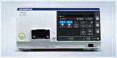
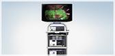
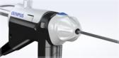
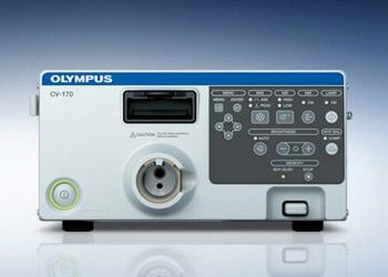
CV-170 Video Processor and LED Light Source
Office Video Processor
CV-170 Video Processor and LED Light Source
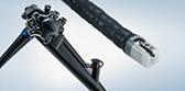
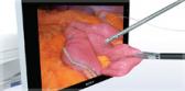
You can only treat what you can see. HD videoscopes have quickly become the standard in many ENT procedures due to the ability to examine mucosal patterns with greater clarity than traditional fiber or SD videoscopes. For the first time ever, HD laryngoscopy is now available to the patients who need it most at the point where it is most valuable: diagnostic laryngoscopy in the office.
With LED light, HD scope support, and Narrow Band Imaging™ (NBI™) Technology built into one, compact box, the CV-170 provides physicians with exceptional diagnostic tools at an affordable price for any office.
Key Benefits
Advanced Imaging
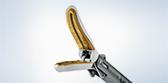
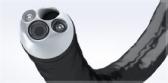
- The CV-170 is compatible with HD videoscopes to provide the image quality of a rigid scope with the comfort of a flexible rhino-laryngoscope.
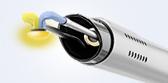
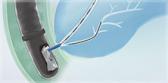
- Patented NBI™ Technology built into the CV-170 enhances visibility of vascular structures on the mucosal surface.
Reduced Costs
- The CV-170’s LED light source reduces cost by virtually eliminating the need to replace expensive Xenon or Halogen light bulbs.
- The platform’s ultra-quiet and power-saving design also uses less energy than previous systems.
Greater Efficiency
- The CV-170’s compact design saves valuable space in procedure rooms.
- The system's backwards compatibility with previous-generation products extends the life of existing scopes and camera heads.
Product Support
Ordering Info CV-170
| Video System & Ancillary Devices | |
| CV-170 | All-in-One HD Video System w/LED & NBI™ Technology |
| MAJ-1981 | Olympus Keyboard |
| MAJ-1925 | Olympus 2GB USB Portable Memory |
| MAJ-438 | Remote Accessory Cable |
| MAJ-1981 | Olympus Keyboard |
| MAJ-1925 | Olympus 2GB USB Portable Memory |
| LMD-2110MD/OL | 21.5" LCD HD Widescreen Medical Grade Monitor |
| USB200 | SD Image/Video Capture to USB Drive |
| IS40950 | nStream DX V9.0 Image and Video Recording with DICOM |
| UP-25MD | Sony Color Video Printer, small format (for office use) |
| TC-C2-PS | Primary Endoscopy procedure cart with power supply and keyboard tray |
| AR-T10E | Coupler |
| AR-T12E Coupler | All-in-One HD Video System w/LED & NBI™ Technology |
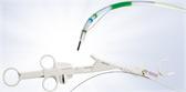
| ENF-VH | HD Rhinolaryngoscope |
| Product Specs | |
| Power Supply | |
| Voltage | 100-240V AC: within +/- 10% |
| Frequency | 50/60 Hz: within +/- 1 Hz |
| Rated | Input 200 VA |
| Size | |
| Dimensions | (W x H x D) 295 x 145 x 425 mm (W x H x D) |
| Weight | 11.0 kg |
| Observations | |
| Examination Lamp | LED Lamp |
| Analog HDTV signal output | Either RGB (1080/60I: NTSC)/(1080/50I: PAL) or YPbPr (1080/60I: NTSC)/(1080/50I: PAL) output can be selected. |
| Analog SDTV signal output | VBS composite (480/60I: NTSC)/(576/50I: PAL), Y/C (480/60I: NTSC)/(576/50I: PAL), and RGB (480/60I: NTSC)/(576/50I: PAL): simultaneous outputs possible. |
| Digital signal output | HD-SDI (SMTPE 292M), SD-SDI (SMPTE 259M) and DVI (WUXGA, 1080p or SXGA) can be selected. |
| White balance adjustment | White balance adjustment is possible using the white balance button on the front panel. |
| Color tone adjustments | Red adjustment: ±8 steps, Blue adjustment: ±8 steps, Chroma adjustment: ±8 steps |
| Automatic gain control (AGC) | The image can be electronically amplified when the light is inadequate due to the distal end of the endoscope being too far from the object. |
| Noise reduction | Noise is corrected by image processing |
| Iris | The auto iris modes can be selected using the "iris mode" switch on the front panel. Peak: The brightness is adjusted based on the brightest part of the endoscopic image. Average: the brightness is adjusted based on the average brightness of the endoscopic image. |
| Image enhancement setting | Fine patterns or edges in the endoscopic images can be enhanced electrically to increase the image sharpness. Either the structural enhancement or edge enhancement can be selected according to the user setup
|
| Freeze | An endoscopic image is frozen using an endoscope or the "FREEZE" key on the keyboard |
| NBI™ Technology Observation | This is one of optical-digital observations using the narrow band observation light |
| Remote control | DVR, Video printer, Image filing system, Flushing pump, Endoscopic CO2 regulation unit can be controlled (specified models only). |
| Documentation | |
| Patient data | Patient ID, Patient name, Sex, Age, Date of birth, Date of recording (time, stopwatch), Comments can be displayed in the endoscopic image screen. |
| Displaying the record state | Portable memory and internal buffer, DVR, Video printer, Image filing system can be displayed on the monitor. |
| Advance registration of patient data | Patient ID, Patient name, Sex and age, Date of birth of up to 50 patients can be registered. |
| Portable Memory | |
| Media | MAJ-1925 (OLYMPUS) |
| Recording Format | TIFF: no compression, JPEG (1/5): approx. 1/5 compression, JPEG (1/10): approx. 1/10 compression |
| Number of recording images | TIFF: approx. 227 images, JPEG (1/5): approx. 1024 images, JPEG (1/10): approx. 2048 images |